


| 상품명 | 3D 아나토미_Superior Orbit |
|---|---|
| 판매가 | 가격 전화문의 |
| 상품요약정보 | 독일제품. 3D아나토미 모델. |
| 국내·해외배송 | 국내배송 |
| 배송비 | 3,000원 (100,000원 이상 구매 시 무료) |
| 수량 |   |
| SNS 상품홍보 |
|---|

 |
(최소주문수량 1개 이상 / 최대주문수량 0개 이하)
사이즈 가이드수량을 선택해주세요.
위 옵션선택 박스를 선택하시면 아래에 상품이 추가됩니다.
| 상품명 | 상품수 | 가격 |
|---|---|---|
| 3D 아나토미_Superior Orbit |


|
가격 전화문의 ( |
| 총 상품금액(수량) : 0 (0개) | ||


3D 아나토미 시리즈는
인체해부학적으로 정확하게 설계되어 실제와 거의 동일하나
카데바에 따르는 윤리적인 문제 없이
합리적인 가격으로 구입가능하며
고객의 요청에 따라 실제보다 크게 혹은 작게 생산 가능합니다
자세한 문의는 전화상담 바랍니다.(문의전화: 031-702-2027)
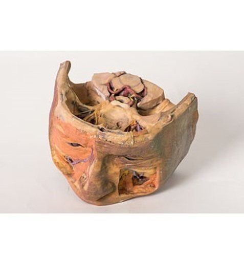
This 3D printed model captures a dissection in which the calvaria and cerebrum have been removed to expose the floors of the anterior and middle cranial fossae. The midbrain has been sectioned at the level of the tentorium cerebelli and on the cross sectional surface one can identify the superior colliculi, cerebral peduncles and the substantia nigra. Anterior to the mid-brain the vertebral artery can be clearly identified rising from the posterior cranial fossa and dividing into the posterior cerebral arteries. Anterior to this in the region of the sella turcica one can identify the internal carotid arteries emerging from the roof of the cavernous sinus medial to the anterior clinoid processes and beneath and lateral to the optic nerves and chiasm. The oculomotor nerves are visible penetrating the roof of the cavernous sinuses on the left and right posterior to the point where the internal carotid arteries emerge.
Anteriorly in the midline of the anterior cranial fossa lies the crista galli with the olfactory bulbs still present above the cribriform plates on either side. On the right the orbital plate of the frontal bone (the roof of the orbit) has been removed to expose the frontal nerve splitting into the supraorbital and supratrochlear nerves lying superior to the levator palpebrae superioris. The trochlear nerve is visible entering the superior aspect of the superior oblique muscle belly on the medial aspect of the orbit. Ethmoidal air cells have been exposed in the medial orbital wall by removal of the part of the lamina papyracea. On the left the levator palpebrae and superior rectus muscles have been divided along with the frontal nerve to expose the optic nerve, nasociliary nerve, ophthalmic artery and superior ophthalmic vein in the intraconal space.
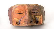
The face has been dissected to show facial muscles around the orbit on the right and the infraorbital nerve on the left. The infratrochlear nerve is also shown on the right and facial veins and arteries are also visible.
교환 및 반품이 가능한 경우
구매하신 제품에 대해 문제가 발생할 경우 제품이 원래 배송한 상태와 동일한 상태에서 구매하신 후 7일 이내라면 언제라도 100% 교환이 가능합니다.
환불은 제품수령 후 3일 이내 환불처리(카드승인 취소 및 온라인 무통장 입금 등) 해 드립니다.
상품의 사용후기를 적어주세요.
게시물이 없습니다


상품에 대해 궁금한 점을 해결해 드립니다.
게시물이 없습니다