


| 상품명 | 1715 Heart internal structures |
|---|---|
| 판매가 | 가격 전화문의 |
| 국내·해외배송 | 국내배송 |
| 배송비 | 3,000원 (100,000원 이상 구매 시 무료) |
| 수량 |   |
| SNS 상품홍보 |
|---|

 |
(최소주문수량 1개 이상 / 최대주문수량 0개 이하)
사이즈 가이드수량을 선택해주세요.
위 옵션선택 박스를 선택하시면 아래에 상품이 추가됩니다.
| 상품명 | 상품수 | 가격 |
|---|---|---|
| 1715 Heart internal structures |


|
가격 전화문의 ( |
| 총 상품금액(수량) : 0 (0개) | ||


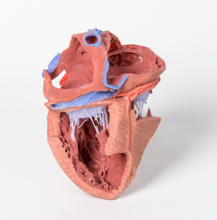
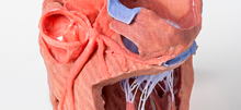
This 3D printed heart has been dissected to display the internal structures of the chambers. At the base of the heart the termination of the superior vena cava is preserved entering the right atrium. Part of the inferior vena cava is also preserved on the inferior aspect of the right atrium; however, most of the vessel lumen and much of the anterior wall has been removed to expose the pectinate muscles of the right auricle and the fossa ovalis (which is nearly translucent in the 3D print). The anterior wall of the right ventricle has also been removed to expose the right atrioventricular valve and its three cusps (anterior, posterior, and septal), including the chordae tendineae connecting them to respective papillary muscles projecting from trabeculae carneae (including a septomarginal trabecula entering the anterior papillary muscle from the interventricular septum). The smooth wall of the conus arteriosus is also exposed leading to the pulmonary semilunar valve (left, right, and anterior cusps) at the base of the pulmonary trunk. Preserved and encircling the right atrioventricular valve is the right coronary artery, ultimately passing to the posterior aspect and the origin of the posterior interventricular artery and atrioventricular nodal artery.
On the posterior side of heart the terminations of the pulmonary veins are visible entering the opened left atrium. Just anterior to the depression of the fossa ovalis in the interatrial septum the left atrioventricular valve with its two cusps (anterior and posterior) is preserved, along with the associated chordae tendineae and papillary muscles in the ventricle. The walls of the opened left ventricle preserve well-developed trabeculae carneae. At the apex of the ventricle the aortic semilunar valve (with left, right, and posterior cusps preserved) can be seen at the base of the sectioned aorta alongside the origin of both coronary arteries. The left coronary artery in this specimen is very short, giving rise almost immediately from its origin to the left anterior descending artery, the diagonal artery, the ramus intermedius, and the circumflex branch. The latter branch passes between the left atrium and ventricle adjacent to the opened coronary sinus leading to the right atrium. The left anterior descending branch penetrates the myocardium in this individual and travels through the tissue, only emerging superficially to become visible again near the apex
교환 및 반품이 가능한 경우
구매하신 제품에 대해 문제가 발생할 경우 제품이 원래 배송한 상태와 동일한 상태에서 구매하신 후 7일 이내라면 언제라도 100% 교환이 가능합니다.
환불은 제품수령 후 3일 이내 환불처리(카드승인 취소 및 온라인 무통장 입금 등) 해 드립니다.
상품의 사용후기를 적어주세요.
게시물이 없습니다


상품에 대해 궁금한 점을 해결해 드립니다.
게시물이 없습니다